What Actually Happens to Your Skin Photos Before a Telehealth Dermatologist Reviews Them
Secure processing and AI checks prepare your photo for a doctor's eyes, not a diagnosis.

AI-assisted teledermatology with mandatory clinician sign-off uses AI to check photos or suggest possible diagnoses, while a physician reviews the case and decides treatment. AI does not replace a confirmatory diagnosis.
Why most patients have no idea what happens to their skin photo after they hit submit
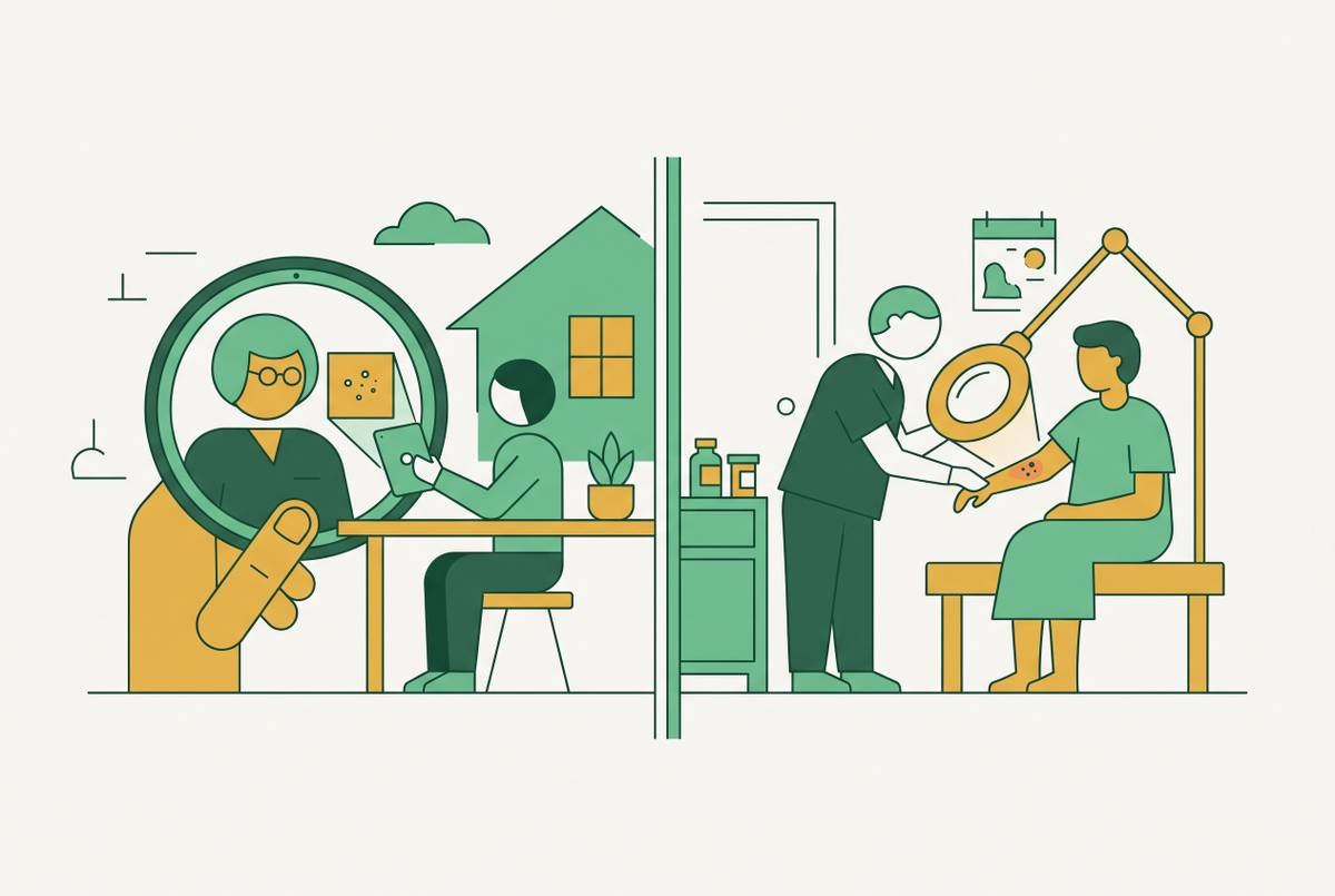
You hit submit, and it feels like your part is done. It is easy to picture a dermatologist opening the photo, looking at the skin, and sending back an answer. What actually happens next? Between upload and review, the image passes through technical processing, privacy safeguards, and, on platforms that use them, AI-assisted quality checks and diagnostic support.
The patient sees little of what happens in those steps. Yet not knowing what they require can lead you to submit a photo that gives the dermatologist too little to work with. The dominant model separates submission from clinical review, so you are not present when the physician examines the image. An answer can take up to a few days, and a missing detail cannot be clarified in the moment as it could during a live visit.
What store-and-forward teledermatology is and why it became the standard model
Store-and-forward, usually shortened to SAF, uses electronically stored information, including photographs and demographic data, for a clinical decision made separately from the patient encounter. The patient or referring physician supplies images and a clinical history, which the dermatologist reviews later. No shared appointment time is needed. That removes the scheduling constraint of live video, while the ability to work on any internet connection speed makes the format practical for more patients.
The need extends beyond convenience. Frazier et al.’s 2025 geospatial study of dermatologists who take part in a public health insurance program found that regions where millions of people lived had no reasonable access to those doctors within a one-hour drive. Patients in rural areas often wait weeks for an appointment. SAF redistributes dermatologists’ attention, allowing a board-certified physician to review cases from underserved areas without either party traveling.
Live video and hybrid care are other ways to get care. Store-and-forward remains the most used, however, because the patient and dermatologist do not have to be available together. That distinction explains the rest of the photo’s journey.
Step one — what the platform does to your photo the moment it arrives
Before a clinician or AI model assesses the skin, the platform processes the upload to protect privacy and prepare it for clinical use. A skin photograph may also reveal who it belongs to through a face, marks that stand out, or tattoos. Such images are protected health information under HIPAA and belong in a patient portal or the platform’s designated upload tool.
Secure transfer is only part of the picture. The photo becomes part of a clinical file rather than a one-time message that loses its purpose after someone reads it. Patients retain the right to access their images as part of the medical record.
What can happen to the image beyond treatment? HIPAA allows images to be used and shared for treatment, payment, and healthcare operations without asking the patient for further authorization. Distribution for education, marketing, or external publication requires a separate HIPAA Authorization for Photography. For direct-to-consumer services outside a traditional provider relationship, however, the terms for how images are stored and shared differ widely, and full HIPAA coverage cannot be taken for granted on every platform. Once the photo is securely in the system, an AI-assisted workflow can begin assessing whether it is useful enough for review.
Step two — how AI quality-checks the photo before a dermatologist opens it
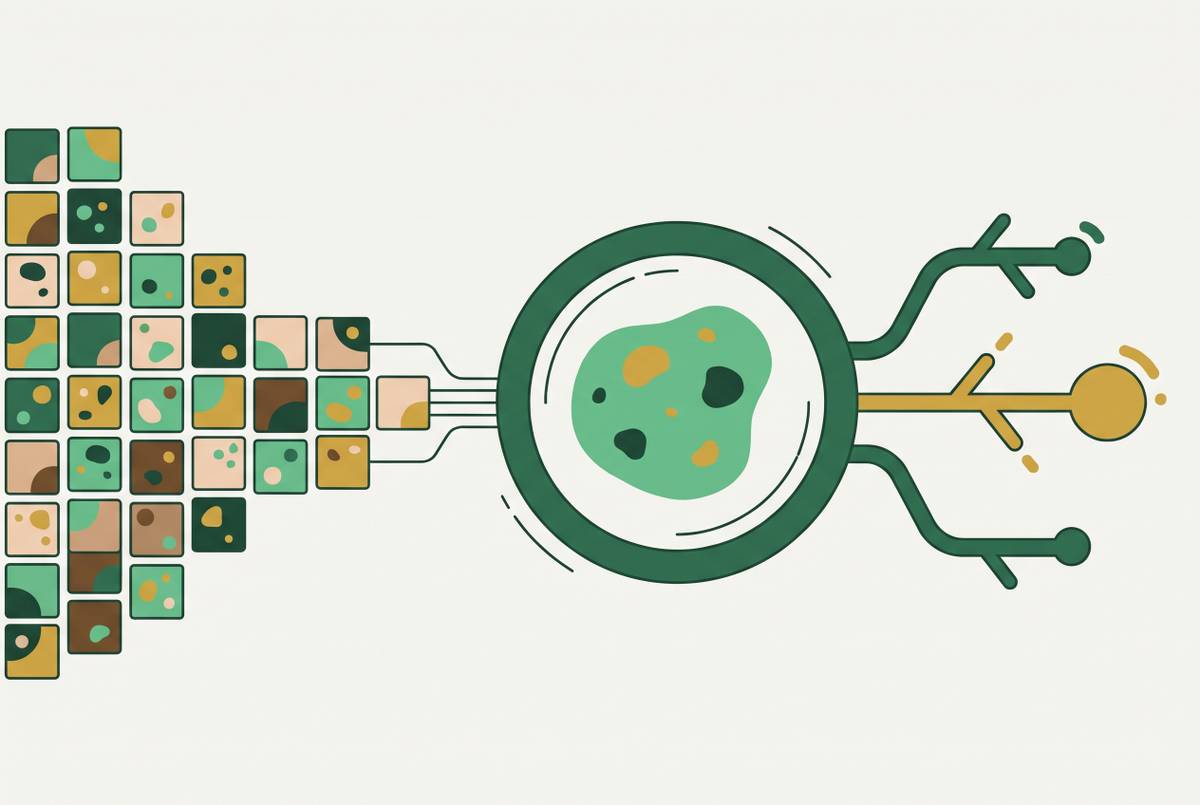
AI checks visibility before considering disease. Image quality assessment tools can flag photographs that are too blurry, too dark, or too poor in other ways for a clinician to use before a dermatologist spends time on them. This is quality triage.
Enhanced Dermatology Image Quality Assessment via Cross-Domain Training describes deep-learning systems including TrueImage, ImageQX, and DIQA in this role within SAF workflows. These examples of image assessment tools do not show that every teledermatology service screens images in the same way. Their job is narrower than it may sound. Passing a quality check means a photograph has cleared a usability hurdle.
Training data limits this layer of AI, too. The same paper introduces cross-domain training that combines dermatology and non-dermatology image datasets, with a dataset named Legit.Health-DIQA-Artificial. The approach addresses the small size of dermatology-specific image quality datasets. It aims to improve assessment of the photograph itself.
A failed check typically means a request to resubmit. That loop can add time and frustration before the physician has even begun the clinical assessment. A better initial photograph can avoid an image-quality problem, but it cannot guarantee that the condition is suitable for remote care. Even after a photo passes, another AI layer may be involved, this time suggesting possible diagnoses for the dermatologist to consider.
Step three — what AI does — and cannot do — as a diagnostic support tool
Instead of assessing whether the image is usable, AI can generate a differential diagnosis listing possible explanations for what the image shows. It can flag and rank those possibilities for the reviewing clinician. The distinction is essential because a useful suggestion is not the same as a confirmed diagnosis.
AI has shown dermatologist-level diagnostic accuracy for skin lesions from clinical and dermoscopic images in controlled settings. Real-world evidence, performance across skin phototypes, and health economic viability remain unresolved challenges. An accuracy claim therefore needs context about the images and patients on which the tool was assessed before it can support expectations about your own submission.
Large dermatology image repositories disproportionately contain lighter skin tones, classified as Fitzpatrick I–III. A 2023 Uganda study that tested a leading AI tool on patients with Fitzpatrick type 6 skin found accuracy of just 17%, far below how well the same model did on Caucasian skin. That result shows why a tool cannot be assumed to work as well across skin tones, though it gives no single score for all dermatology AI. It also complicates the access argument, because people who could benefit from remote care may be poorly represented in the data used to train its tools.
There is useful clinical potential as well. FotoFinder’s Moleanalyzer pro has drawn praise for finding skin cancer early, while clinicians stress that it should help them use their expertise without taking their place. In a workflow with mandatory clinician sign-off, a board-certified dermatologist reviews the case and makes the treatment decision, rather than treating an AI ranking as approval to prescribe.
That distinction does not establish which service is “best.” An image quality tool, a diagnostic support product, and a telehealth platform with a physician approval requirement are different things. Naming one does not establish the policies of another. AI findings remain inputs to a licensed physician’s judgment.
Step four — what does the doctor review before approving treatment?
By the time the dermatologist opens the submission, there is more than a photograph on the screen. The clinical package includes the image, the patient’s history and symptom description, and any AI-generated flags. Each part supplies something the others cannot. A sharp image can show appearance, while the written account explains when the problem began and how it has behaved.
Patient-submitted images tend to have lower and more variable quality than clinician-taken photographs, with more variable agreement between remote and in-person diagnoses. Poor photographs are one cause of that disagreement, but not the only one. Missing dermoscopy for pigmented lesions, a need for palpation, or incomplete photographs of a condition’s distribution across the body can also leave important gaps. An AI quality pass cannot resolve those gaps.
Acne has shown the highest diagnostic match across image types, while suspected melanoma, nonmelanoma skin cancers, and undiagnosed pigmented lesions require in-person evaluation regardless of photo quality. That reflects a limit of the format. A dermatologist can assess what the submission supports while recognizing that the next decision requires an examination or a different kind of image.
Most studies of SAF for skin cancer show that diagnoses often agree with those made in person, with teledermoscopy improving results and dermoscopy bringing accuracy up to 80% for skin cancer cases. Dermoscopy is available to clinicians. Those results therefore should not be treated as a promise about a phone photo uploaded from home.
Mandatory sign-off gives the clinician responsibility for interpreting these limits. It does not make every submission sufficient for treatment. The physician’s endpoint may be a clinical decision based on the supplied information or a request for in-person evaluation. What arrives on the screen sets the bounds of that judgment.
What patients can control — how to submit photos that survive every step of the pipeline
Prepare both the photographs and the written history. Survey data found that evaluators felt comfortable making medical decisions for only 55.1% of patient-submitted images. You can improve the inputs without controlling the diagnosis.
Start with natural daylight rather than flash, fluorescent light, or dim indoor lighting. Light is the highest-impact variable in the submission. It affects how true the photo is to the skin’s tone and texture, which shapes both the AI quality check and how the dermatologist judges the image. Avoiding glare and color distortion helps the image show the skin accurately.
Give the dermatologist more than one view. The photographs should include an overall photograph, a mid-range shot showing the lesion in context, and a close-up, using multiple angles. Include an object for scale, with a coin as the clinical standard, and tap the lesion on your phone screen to lock focus before shooting. If you are monitoring a condition over time, use the same angle and distance each time so that changes can be judged across comparable images.
The written history deserves the same care as the photos. Describe when the problem began, what makes it better or worse, and any treatments already tried. Include triggers that may have brought on the problem, such as sun exposure, hormonal changes, new products, or stress. Those details give the clinician context that the image cannot supply, even when focus and lighting are good.
Patient education helps make these steps a regular part of care. Saade et al.’s 2025 randomized cohort study highlights its value in improving image quality and diagnostic outcomes, because patients commonly photograph their own lesions without guidance. Platforms can support that work by prompting for context, checking image quality before submission, and explaining what the clinician can and cannot assess.
Finally, treat an in-person referral as part of care. Conditions requiring palpation or dermoscopy, and lesions the clinician flags for examination, should move to an in-person appointment without delay.
Sources
- Teledermatology Diagnostic Accuracy: A Randomized Cohort Study Comparing Three Image Acquisition Techniques - Saade - 2025 - International Journal of Telemedicine and Applications - Wiley Online Library
- Enhanced Dermatology Image Quality Assessment via Cross-Domain Training
- Store-and-Forward Teledermatology for Assessing Skin Cancer in 2023: Literature Review