Mole Changes That Warrant a Dermatologist Visit
Early detection of melanoma raises five-year survival rates from 34% to over 99%.
Melanoma diagnoses in the country covered by this data climbed 42% between 2015 and 2025, according to American Cancer Society data. That sounds like bad news, and in some ways it is. But death rates haven't climbed anywhere near as fast: people are catching this disease earlier than they used to.
About 2.2% of Americans will get a melanoma diagnosis at some point in their life. As of 2023, an estimated 1,573,288 people in the U.S. were living with it. Those are big numbers. If there's a mole on your arm you keep glancing at, 76.9% of melanoma cases, per SEER data, get caught while the disease is still local, still contained, still highly treatable. Most people do catch it in time. They just know what to look for first.
This piece covers that knowledge: the ABCDE framework dermatologists use to describe which mole changes deserve a real look, and why "evolving" (the E) drives the decision to check a mole more than the other four letters combined.
What survival data actually says about the value of catching changes early
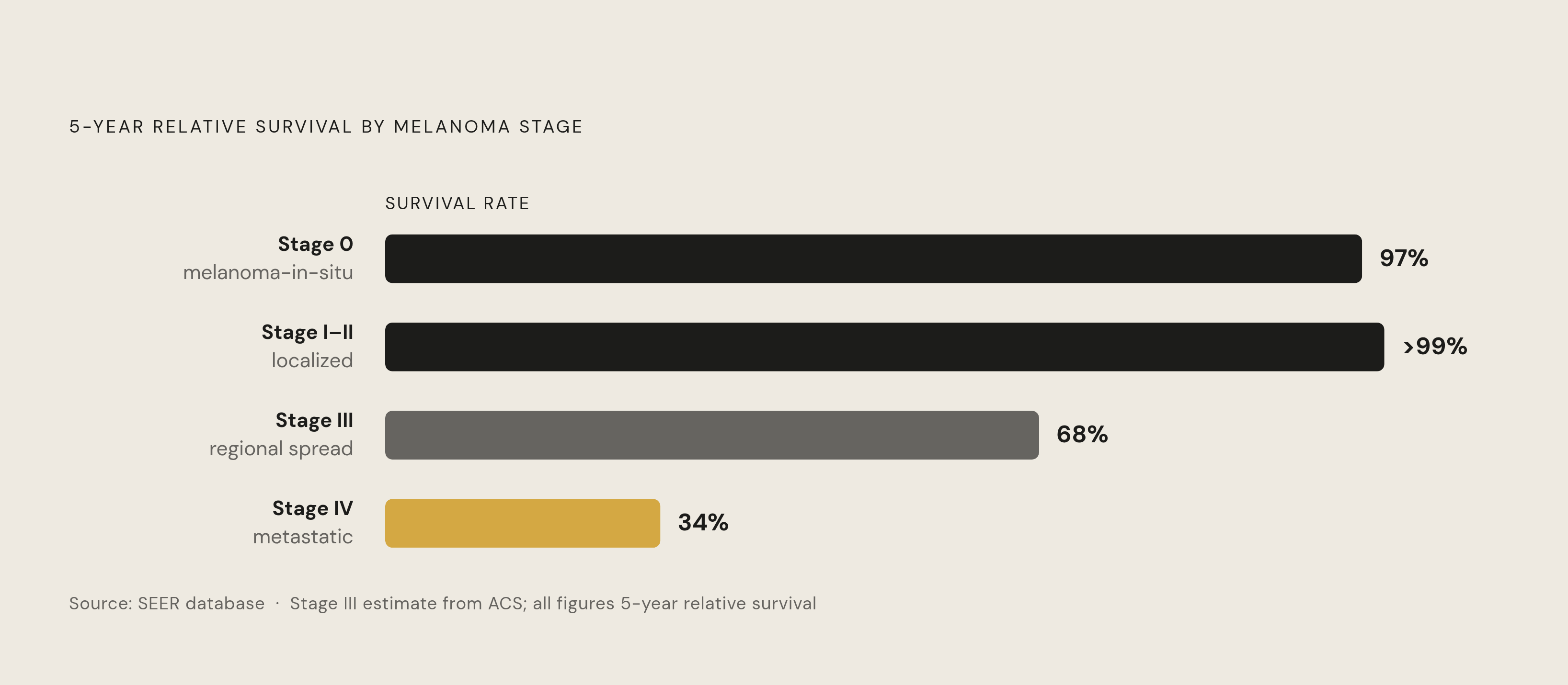
Start with the gap, because the gap is the whole argument. Five-year relative survival for localized melanoma exceeds 99%. Once the disease has spread to distant sites, that number falls to somewhere around 34 to 35%, based on SEER figures. Same disease, wildly different outcome, and the difference comes down almost entirely to timing.
Break it down by stage and the pattern holds. Stage 0, or melanoma-in-situ, carries a 97% five-year survival rate. Stage IV is around 30%. Framed differently: 99 out of 100 people survive at least five years when melanoma is caught before it reaches the lymph nodes, according to data cited by GoodRx.
None of this means survival is purely a function of vigilance. Treatment has genuinely improved. Checkpoint inhibitor immunotherapies have more than doubled survival for advanced-stage patients over what earlier treatments achieved. So it's not that stage IV used to be a death sentence and staying alert makes it disappear. It's that earlier stages still offer more options, simpler treatment, and dramatically better odds, no matter how good later-stage treatment gets.
Which raises the obvious next question: if timing matters this much, how does a person actually know when a mole has crossed the line from ordinary into worth checking? That's what ABCDE was built to answer.
What a mole actually is and why some are more likely than others to become problematic
A mole, or nevus, is a benign cluster of pigment-producing cells called melanocytes. Most people aren't born with the moles they have as adults. They show up during childhood and early adulthood, and new ones can keep appearing into early adulthood.
Melanocytes originate from the neural crest during embryonic development. That's part of why melanoma, despite starting in the skin, can behave differently than other skin cancers and occasionally show up in places far from any mole at all. It's a cancer of a specific cell type, not just a location.
The mechanism behind sun exposure risk is straightforward once you see it laid out. UV radiation damages DNA inside skin cells. The immune system repairs a lot of that damage, but not all of it. Damage accumulates over years, and eventually a cell picks up the wrong mutation and starts dividing without the usual brakes. That's the biological chain connecting a childhood sunburn to a diagnosis decades later.
Dysplastic nevi complicate the picture. These are moles larger than average, often carrying more than one shade (light brown, dark brown, sometimes pink), and they tend to run in families. They also carry a higher lifetime melanoma risk on their own. Why does this matter for self-exam? Because a dysplastic nevus can look like it's failing the ABCDE test even when it's stable and has looked that way for years. Context matters as much as the checklist.
There's also a genuine scientific debate: does melanoma arise from within an existing mole, or does it develop in the skin next to one? Researchers haven't settled this. Practically, it means the skin around a mole deserves attention too, not just the mole itself.
A few factors lower the threshold for concern meaningfully:
- More than 50 ordinary moles raises risk; cross 100, and risk climbs roughly sevenfold
- Five or more atypical moles increases risk, and the risk rises further with each additional one
- Family history matters: roughly 1 in 10 people with melanoma have a family history of the disease
- Congenital moles, present at birth, are also considered a risk factor worth noting
- A history of blistering childhood sunburns or indoor tanning use
- A personal history of melanoma or other skin cancer
One disparity deserves direct mention. Melanoma is less common overall in people with highly pigmented skin, but acral melanoma, the kind that shows up on palms, soles, and under nails, doesn't discriminate by skin tone. And the outcome gap is stark: Black patients see roughly 70% five-year survival compared to 94% for White patients. That gap traces mostly to later-stage diagnosis, not a more aggressive disease. It's a detection problem, and detection problems are exactly what a framework like ABCDE is meant to solve.
How to read Asymmetry and Border as a pair, not as standalone checkboxes
Picture drawing a line through the middle of a mole. If one half doesn't mirror the other, that's asymmetry, the "A" in ABCDE. Benign moles tend to be symmetrical. But symmetry alone doesn't clear a mole either; plenty of early melanomas still look fairly even.
Border is the companion signal. Benign moles usually have smooth, well-defined, rounded edges. Melanoma borders tend toward ragged, notched, or scalloped, sometimes with edges that appear uneven or indistinct rather than stopping cleanly. That bleeding-edge look stands out on its own.
Here's the caveat: asymmetry and irregular borders are warning features, not diagnoses. A border you can see with your eyes tells you nothing about how far abnormal cells have spread beneath the surface. These two criteria carry more weight when they're new, when they're changing, or when they show up alongside other signs, not in isolation.
Good lighting helps here more than people expect. A hand mirror for spots on the back or scalp, decent overhead light, maybe a phone flashlight for shadowed areas. This is really the mechanical starting point for a proper self-exam, something worth building into a monthly habit rather than treating as a one-time inspection.
Structure is one layer of the picture. Color is often where change becomes visible first, sometimes before shape does anything at all.
What color variation in a mole signals and why a single shade matters
A typical benign mole holds one shade, brown or tan, evenly through the whole lesion. The moment a mole starts showing more than one color inside it, that's worth attention.
What does suspicious color variation actually look like? Multiple shades of brown mixed with black, red, white, or gray, all inside the same mole, according to sources including Cleveland Clinic. Not a mole that's simply dark. A mole that's dark in one spot and pink in another and maybe grayish along one edge.
Color, like border, can't stand alone as proof of anything. It can't tell a clinician how deep a lesion has grown, and it can't by itself distinguish melanoma from a perfectly harmless spot. This is the cumulative logic of ABCDE: no single letter convicts, but each one raises the case for a closer look.
Here's a concrete version of what this looks like in practice. A mole that's been uniformly brown for years suddenly develops a darker patch in the center, or a reddish tinge along one side. That kind of shift, the change itself, is what warrants a call to a dermatologist, not just the current appearance on its own (a point emphasized by sources like PS Dermatology). Some sources go as far as calling color one of the most clinically useful indicators in day-to-day practice, though that shouldn't be read as a guarantee of anything either way.
Why diameter is the most misunderstood part of ABCDE
The number most people remember from ABCDE is 6 millimeters, roughly the size of a pencil eraser. The American Academy of Dermatology uses that threshold as a flag.
But treating 6mm as a safety line is where people get this wrong. Early melanomas can be smaller than that. A lot smaller. Size alone was never meant to rule anything out; it's a flag above the line, not a green light below it.
What determines the next step is not the number on the ruler but direction. Is the mole getting bigger? At what pace? A mole that's grown noticeably over a few months deserves attention regardless of whether it's crossed 6mm yet.
Nodular melanoma makes this point sharply. It's an aggressive subtype that can appear suddenly as a firm, raised bump and grow fast, over a matter of weeks rather than years. A single measurement taken at one moment in time can be misleading if nobody's tracking how quickly that measurement is changing.
The practical fix is simple: measure with a small ruler, or better, take photos over time and compare them side by side. Cleveland Clinic's self-exam protocol recommends exactly this, photographing moles specifically to make future comparison possible. One look tells you almost nothing. A series of looks tells you everything.
Why "evolving" is the criterion that does the most work
Ask which single letter in ABCDE carries the most weight, and the Bare Dermatology source calls it the most critical criterion: evolving. That's not editorial spin, that's a clinical framing straight from the source material.
What counts as evolving? Any change in size, shape, color, or elevation. New symptoms showing up where there weren't any before, itching or scabbing per Cleveland Clinic, bleeding or crusting per PS Dermatology. A mole that used to sit flat against the skin starting to raise up. One that begins itching for no obvious reason.
Why does this catch what the other four letters sometimes miss? Because a melanoma can be actively developing before it shows clear asymmetry, before its border turns ragged, before it picks up a second color, before it grows past 6mm. Change is often the earliest visible signal, arriving before the static snapshot criteria have anything to show.
Some melanomas don't produce a change the patient notices at all. Absence of perceived change doesn't clear a mole. That's exactly why self-exam works best as a complement to professional evaluation, not a replacement for it. A dermatologist's trained eye and dermoscope catch things a mirror check won't.
And the timeline can move faster than most people assume. Melanoma can develop over weeks to months, not exclusively over years, and the more aggressive subtypes can shift noticeably within a matter of weeks. So the instinct to wait another month and "see if it changes more" isn't really a neutral choice once change has already been spotted. It's a choice to spend time that might matter.
Patients who track their skin with some system (photos, a monthly check, a ruler kept in a drawer somewhere) walk into a dermatology appointment able to describe change with precision. That precision speeds up the whole evaluation. Telling a clinician "this started as a small dark spot eight weeks ago and now has a raised edge" gets you further, faster, than "it looks weird now."
What the "ugly duckling" sign catches that ABCDE doesn't
Here's a sign that doesn't show up on the ABCDE checklist at all, and PS Dermatology treats it as its own category: the ugly duckling sign. A mole that looks distinctly different from every other mole on someone's body, even if it doesn't technically break any single ABCDE rule, deserves a second look.
Why does this work as a signal? Because most people's benign moles follow a personal pattern, similar size, similar color family, similar texture, almost like a signature. A mole that breaks from that pattern stands out precisely because it doesn't belong, not because it's obviously abnormal in isolation. One might argue this makes ugly duckling more intuitive than ABCDE for everyday self-checks. It doesn't require memorizing five criteria, just a sense of what looks out of place.
Dermatologists use this concept alongside ABCDE, not instead of it. It catches lesions that would look perfectly ordinary on someone else's skin but look wrong on this particular person's skin.
Beyond ABCDE and ugly duckling, a handful of other signs justify a visit on their own:
- Bleeding that happens on its own, or with barely any contact at all, since melanomas often carry fragile, abnormal blood vessels
- Itching, tenderness, or pain that keeps coming back or won't explain itself, especially paired with visible change
- A mole sitting somewhere hard to monitor, like the scalp, between the toes, under a nail, or across the back
- A new mole showing up after age 30
- A dark band in a fingernail or toenail that's widening or changing, or pigment spreading onto the skin around the nail
- A sore or spot that simply won't heal after a few weeks
None of these symptoms diagnose melanoma by itself. Plenty have completely benign explanations. But that's exactly the point: they're reasons to get a professional look, not reasons to self-diagnose one way or the other.
How to do a skin self-exam that will actually surface changes worth acting on
A monthly check is the commonly suggested interval, per Advanced Dermatology, P.C., though it's worth being honest that this isn't a proven universal rule so much as a practical, workable standard.
Cleveland Clinic lays out a specific sequence to follow step by step. Start in a well-lit room with a full-length mirror and a hand mirror for backup. Check the front and back of the body, then raise the arms to get the sides. Look inside the mouth. Move down the forearms and upper arms, check the palms, check between the fingers. For areas that are genuinely hard to see, the back, the buttocks, the genital area, use the hand mirror and take the time. Lift the breasts and check underneath. Work down the backs of the legs, the soles of the feet, between the toes. Finish with the scalp and neck, parting hair section by section or using a blow dryer to get a clear view, and if that's not workable alone, ask a barber or hairdresser to flag anything unusual.
Documentation turns this from a vague impression into usable data. Photograph moles so there's something to compare against next month. Keep a small ruler nearby for diameter. Note the location, what it looks like, and the date. None of this is about diagnosing anything at home.
The actual goal of self-exam is narrower than people think: it's to generate information, what changed, when, how fast, that makes a dermatology visit sharper and more useful. A patient who says "this mole used to be one uniform color and now has a darker patch in the center" gives a clinician something concrete to work with. "It looks different" doesn't.
When to make the appointment and what to expect when you do
Certain signs call for a prompt appointment, not a few more months of watching:
- Any ABCDE criterion that's new or has changed noticeably
- A mole that bleeds, crusts, ulcerates, or won't heal without an obvious cause
- Itching, tenderness, or pain that persists or can't be explained, alongside visible change
- An ugly duckling mole, one that just doesn't match the pattern of the others
- Any change that shows up within weeks rather than building slowly over years
A mole evaluation itself is usually quick. It typically starts with a visual exam, often followed by dermoscopy, a specialized magnifying tool dermatologists use to look at pigment patterns beneath the surface. If something looks concerning enough, a biopsy follows. The whole process tends to move fast, and it produces answers in a way self-exam simply can't.
For adults without symptoms or elevated risk factors, the question of how often to pursue formal screening is best discussed directly with a clinician. That's not an argument against getting checked. It means frequency should scale to individual risk factors and a clinician's judgment, not follow one blanket rule for everyone.
For anyone carrying elevated risk, family history, a high mole count, prior atypical moles, immunosuppression, a history of indoor tanning, the threshold for getting something looked at should sit lower. Waiting for two or three ABCDE criteria to stack up before making a call isn't the right instinct for this group. One credible change is enough reason to ask.
And for a lot of everyday skin concerns, a telehealth visit works as a reasonable first step, a way to get a professional opinion quickly and figure out whether an in-person exam or biopsy is the right next move.