Skin Rash Patterns That Signal Systemic Conditions
Recognizing rash patterns can reveal lupus, dermatomyositis, and other systemic diseases.
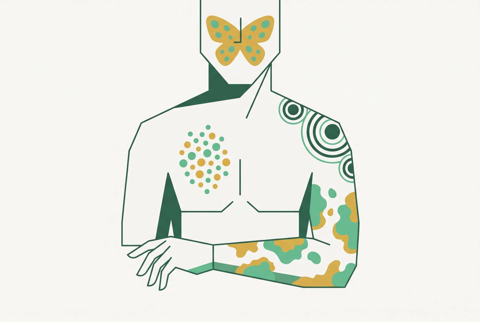
A rash's location on the body, and how it's distributed across it, tells a clinician more than most patients realize. Its location on the body, whether it's symmetrical, and whether it follows sun exposure or a single nerve path: these details separate a rash that's purely local from one that's flagging something happening inside the body. Learning to read those patterns doesn't just satisfy curiosity. It changes how a patient describes what they're seeing, and it can change how fast the right care happens.
Start with the broad rules clinicians actually use. A rash that's widespread and symmetrical is often associated with systemic disease. One confined to sun-exposed skin may suggest photosensitivity. A rash tracking a single strip of skin, following one nerve root, can be consistent with herpes zoster. When multiple distinct sites are affected together, contact dermatitis, something touched the skin and caused a reaction, is one pattern clinicians consider. Involvement of sebum-rich areas such as the scalp, central face, and skin folds may point toward seborrheic dermatitis instead.
Drug eruptions complicate all of this, because they can resemble a wide range of inflammatory skin conditions. These appear fast: widespread, symmetrical, red, and often accompanied by systemic symptoms such as malaise or fever. Those constitutional symptoms are what push a case toward "this is systemic" rather than "this is topical." A patient who says "it's everywhere and it looks the same on both sides" is handing a clinician a completely different diagnostic thread than one who says "it showed up right where my watch band sits." When a rash does suggest systemic involvement, blood work checking organ function and inflammatory markers is the standard next step, not something to put off.
The malar rash and its relatives: what lupus patterns look like and what they mean
Systemic lupus erythematosus (SLE) is a multi-organ autoimmune disease, and its skin findings aren't a side note. They're written directly into the formal diagnostic criteria: four of the eleven criteria set out by a major professional rheumatology body involve skin or mucous membranes, covering malar rash, discoid rash, photosensitivity, and oral ulcers.
Three distinct skin patterns map to different phases of the disease. Acute cutaneous lupus produces the "butterfly" or malar rash, redness across the cheeks and the bridge of the nose that spares the folds beside the nostrils. It's the most recognizable pattern, and probably the one most people picture when they hear the word lupus. Subacute cutaneous lupus looks different: ring-shaped or scaly, psoriasis-like lesions that appear wherever sunlight reaches. Chronic cutaneous lupus, also called discoid lupus, is different again: scarring, plugged hair follicles, thinning skin, concentrated on the face, ears, and scalp, and notably not arranged in a butterfly shape. Discoid lupus also carries a lower likelihood of progressing to full systemic disease.
A 2025 retrospective study looking at 430 SLE patients treated between 2018 and 2023 found malar rash was the single most common lesion type, at 24%. Bullous and subacute rashes followed, each around 19.8%, with discoid rash close behind at 19.1%. No one pattern dominates the picture. An earlier cohort of 150 patients found non-specific findings mattered too: non-scarring hair loss in 86.67% of patients, oral ulcers in 56.67%, vasculitic lesions in about a third. These don't define the diagnosis on their own, but they track disease activity, appearing during flares.
Roughly a quarter of SLE patients have skin involvement as their first presenting sign, and somewhere between 5% and 10% of people diagnosed with cutaneous lupus alone go on to develop systemic disease later. A skin-only diagnosis today doesn't close the door on systemic involvement tomorrow.
When palpable purpura (raised red or violet bumps from small-vessel vasculitis) or livedo reticularis (a lace-like, net-shaped redness on the skin) appears in a lupus patient, it generally means disease activity is climbing, not just that the skin is acting up in isolation.
Rashes that reveal muscle and lung disease: dermatomyositis patterns
Dermatomyositis offers maybe the clearest example of skin as an early warning system. The rash frequently appears before any muscle weakness is clinically obvious, and often before there's any detectable organ involvement at all. That makes the skin the earliest available clue, not a late-stage confirmation.
A few named signs are specific enough to be genuinely useful, so they're worth knowing by name. Heliotrope rash: a purplish-red discoloration across the eyelids, a location that's highly telling on its own. Gottron's papules: flat-topped, violet bumps sitting over the knuckles. Gottron's sign: redness over the elbows, knees, and other extensor surfaces (the parts of a joint that straighten out, as opposed to the crease side). Shawl sign: redness spread across the upper back and shoulders in the shape its name suggests. A careful hand exam might also turn up dystrophic cuticles or abnormalities in the tiny blood vessels at the nailfold.
Why does this matter beyond cosmetics? Because dermatomyositis skin findings predict risk for interstitial lung disease, and in adults, they raise the odds of an underlying malignancy. The rash isn't the endpoint, it's a window into what else might be going on. Muscle involvement tends to follow or run alongside it: weakness and soreness concentrated in the shoulders, hips, neck, and upper arms, the proximal muscle groups closest to the trunk.
Treatment options remain limited. A 2024 systematic review covering 91 studies and 569 patients found no FDA-approved targeted therapy exists for dermatomyositis as of that review. Rituximab was the most commonly used systemic biologic, appearing in 68.3% of biologic use, followed by infliximab at 16%, etanercept at 5.6%, and abatacept at 3.6%. Given how limited the treatment landscape still is, catching the pattern early through the skin carries real weight.
Psoriasis, nail findings, and the joint disease hiding in plain sight
Classic plaque psoriasis has a distribution that's fairly predictable: sharply bordered, silvery-scaled plaques on the elbows, knees, lower back, scalp, and belly button. Notice what's on that list: extensor surfaces and midline structures, not the folds and creases where contact dermatitis or seborrheic dermatitis tend to cluster.
But what if the skin isn't the whole story? About 10% of people with psoriasis go on to develop psoriatic arthritis, joint pain, swelling, and stiffness that often occurs asymmetrically, hitting individual fingers, toes, or the spine unevenly rather than mirroring both sides of the body.
The nails are the bridge between the two. Nail psoriasis, showing up as oil-drop spots under the nail, pitting, lifting of the nail plate (onycholysis), or thickened, misshapen nails, appears in somewhere between 60% and 90% of people who have psoriatic arthritis. That's a wide range, but the direction of the signal is consistent: nail changes alongside skin plaques should prompt a conversation about joints, not just another round of topical steroid.
So the reader managing "just a skin condition" with moisturizer and topical treatment, who also happens to notice stiff or swollen fingers most mornings, isn't describing two unrelated problems. That's one systemic picture, and the nail and joint findings are what changes the clinical calculus from cosmetic management to something that needs a rheumatology referral.
Erythema migrans and why the classic bull's-eye description misleads more than it guides
Lyme disease, caused in the United States by the bacterium Borrelia burgdorferi, moves through stages: early localized, early disseminated, and late disease. Left untreated, it can reach the nervous system, the heart, and the joints. Catching it early matters enormously, which is exactly why the popular image of what the rash looks like deserves scrutiny.
Everyone's taught to look for a bull's-eye. That's the problem. In the United States, only about 20% of Lyme rashes actually show that classic target pattern, compared with roughly 80% in Europe, a geographic gap most patients never hear about. A published survey found that 72.7% of respondents could correctly identify the classic bull's-eye. But when shown the four nonclassic presentations, the correct identification rate averaged only 20.5%. People are good at spotting the version they were taught and bad at recognizing everything else.
And erythema migrans itself, the expanding red rash associated with Lyme, appears in roughly 80% of infections overall, but it can look like a solid red oval, a blotchy uneven patch, or even a cluster of small blisters. Not every case looks ringed. That leaves a meaningful minority of cases where no erythema migrans appears at all. A clean skin exam doesn't rule Lyme out.
Rash mimicry adds another layer of confusion. A published case report described a 76-year-old woman with erythema migrans who also had a heliotrope rash and a shawl sign, features that looked, on the skin alone, like dermatomyositis. Serological testing confirmed Lyme disease instead. Treatment with IV ceftriaxone and doxycycline resolved the erythema migrans and improved her muscle weakness. Two very different systemic diseases, overlapping on the skin, nearly pointing the workup in the wrong direction.
What should actually guide suspicion? Location (an expanding patch starting from a tick bite site), timeline (showing up days to weeks after time outdoors), and constitutional symptoms like fever, fatigue, and joint aches, matter as much as, if not more than, the exact shape of the rash.
Palpable purpura, dermatitis herpetiformis, and rashes that point to organ systems most patients wouldn't guess
Cutaneous vasculitis has a signature finding: palpable purpura, red, purple, or almost black spots and patches that don't fade when pressed. Other forms appear as hive-like plaques, nodules under the skin, or ulcers. The "doesn't blanch" detail isn't trivial. It's the single most useful thing a patient can check at home.
Urticarial vasculitis, one variant, often comes paired with fever, joint aches, and elevated inflammatory markers on lab work. Roughly half of urticarial vasculitis cases are linked to low complement levels and to SLE. That means palpable purpura is never a "wait and watch" rash. Constitutional symptoms, gastrointestinal complaints, blood in the urine, and any lesion that's turning ulcerative or necrotic are the flags that move a case from routine monitoring to urgent systemic workup.
Dermatitis herpetiformis tells a different but equally underappreciated story. It appears as intensely itchy, small blistered bumps concentrated on the elbows, knees, buttocks, scalp, and symmetric and favoring extensor surfaces. It is closely associated with celiac disease. Someone could have this exact rash, with this exact distribution and itch level, and have no obvious digestive symptoms whatsoever, yet the underlying condition can drive the rash without producing noticeable gut-level warning. The skin, in this case, is doing the work the gut symptoms failed to do.
Scleroderma involves progressive tightening and hardening of the skin, especially on the hands and face. It can be associated with internal organ involvement, and skin changes may appear early in the disease course.
A less common but still notable category is rash patterns with no obvious local cause that resist ordinary treatment and may warrant a broader systemic workup. Any rash with no obvious local cause, one that resists ordinary treatment and won't explain itself, is a reasonable argument for a broader systemic workup rather than another round of moisturizer.
How to describe a rash so a clinician (in person or remote) can actually use the information
Five things matter most, and most patients undersell at least two of them.
Location and distribution: which body parts, whether both sides match, whether it follows sun-exposed skin, skin folds, extensor surfaces, or a single narrow band. Morphology, described in plain words: flat or raised, blistered, scaly, purplish, and whether it fades or stays put under pressure. Timeline: when it started, whether it's spreading, and what it looked like at day one compared to now. Associated symptoms: joint pain, muscle weakness, fatigue, fever, gut trouble, vision changes, the constitutional context that turns a rash that looks local into one that clearly isn't. Exposure and triggers: a recent tick bite, time in the sun, a new medication, a dietary change, or a known autoimmune condition in the patient's history.
Why does the blanch test get singled out? Pressing on a rash and watching whether the redness fades is a simple check: redness that persists under pressure suggests blood has escaped the vessels, which is the defining feature of palpable purpura. Palpable purpura doesn't blanch, and that one observation alone is often enough to justify moving faster.
Photographs help more than most people expect, but only if taken well. Natural light, something in the frame for scale, and a shot that captures the full distribution rather than a tight close-up on one bump, gives a clinician reviewing the case remotely far more to work with than a description alone ever could.
That matters more now that message-based, clinician-supervised dermatology consultations have become an available option for many people, especially for triage questions like "does this need an in-person visit this week, or can it wait for a scheduled appointment." Those consultations are only as good as what gets submitted. Distribution, morphology, timeline, associated symptoms: none of that is busywork. It's the raw material the clinician on the other end is actually working from.
And some patterns shouldn't go through a remote consultation at all. Palpable purpura that doesn't blanch, a rash spreading fast alongside fever or joint pain, blistering across a large area of skin, or any neurological symptom showing up alongside a rash: these call for in-person evaluation or emergency care as the first move, not the second.